Table of Contents
Washington University Experience | VASCULAR | Aneurysm - Saccular | 6A3 Aneurysm, basilar tip, ruptured (Case 6) 2
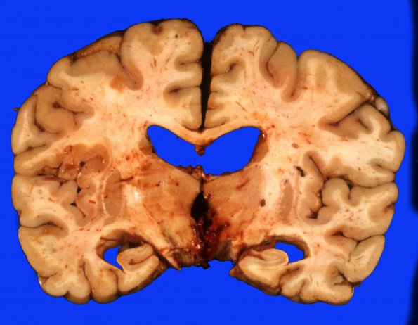
6A3,4 There is intraparenchymal necrosis and hemorrhage in the lower midbrain, primarily on the right side. Serial cross sections of the brainstem and cerebellum at 3-4 mm intervals show blood within the aqueduct of Sylvius and fourth ventricle. There is intraparenchymal \hemorrhage and necrosis in the right pons. An area of dissecting hemorrhage is present in the midbrain. Sections of the brain show evidence of recurrent basilar artery aneurysmal bleeds and terminal rupture. Sections of the basilar artery show a focally ruptured aneurysm in part composed of thrombus, fibrin, macrophages and extensive acute and chronic inflammatory cells. There is both fresh hemorrhage and older hemosiderin deposition. The acute rupture has led to subarachnoid hemorrhage and midbrain and pontine intraparenchymal dissection of blood. There is intraparenchymal necrosis with macrophages, inflammation, and hemorrhage. A non-ruptured aneurysm was found in the right pericallosal artery. Sections of the aneurysm show a thin, dilated artery with areas of atherosclerosis, inflammation, and hemorrhage. ---- The patient clearly suffered diffuse hypoxic damage prior to the terminal event. There is a marked depletion of hippocampal neurons and a subsequent reactive astrocytosis. Sections of the cerebral cortex, cerebellum, and caudate also show a decrease in neuronal density and reactive astrocytes.

