Table of Contents
Washington University Experience | VASCULAR | Cavernous Angioma | 18C4 Cavernous Angioma (Case 18) 12
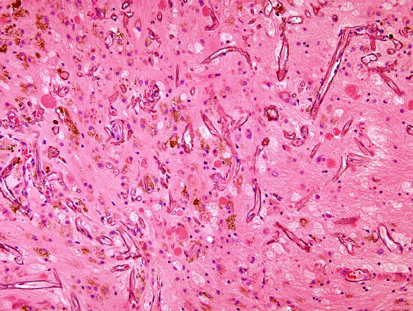
18C4,5 There are patches of small vessels with calcified walls surrounded by hemosiderin stained astrocytes, gliotic parenchyma, and granular axonal spheroids (arrowheads, 18C5) (H&E). These vessels are different in character from those in the malformation itself and may represent reaction to the hypoxic environment of the adjacent brain parenchyma.

