Table of Contents
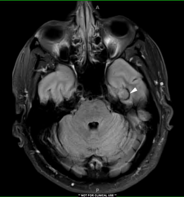
Washington University Experience | VASCULAR | Cavernous Angioma | 1A1 Cavernous Angioma (Case 1) FLAIR copy - Copy
Case 1 History ---- The patient is a 48-year-old man with a seizure disorder and a left temporal lesion. Operative procedure: Left middle fossa craniotomy and lesion resection. ---- 1A1-3 MRI examination demonstrates a cavernous angioma using FLAIR (arrow, 1A1), T1-weighted with contrast (1A2) and T2-weighted with contrast (1A3) sequences are shown. The dark rim (“ferruginous penumbra”) around the T2-weighted MRI scan corresponds to hemosiderin deposition in the adjacent brain tissue.

