Table of Contents
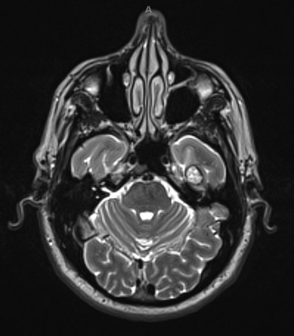
Washington University Experience | VASCULAR | Cavernous Angioma | 1A3 Cavernous Angioma (Case 1) T2 with contrast 2 - Copy
MRI examination demonstrates a cavernous angioma using T2-weighted with contrast sequence. The dark rim (“ferruginous penumbra”) around the T2-weighted MRI scan corresponds to hemosiderin deposition in the adjacent brain tissue.

