Table of Contents
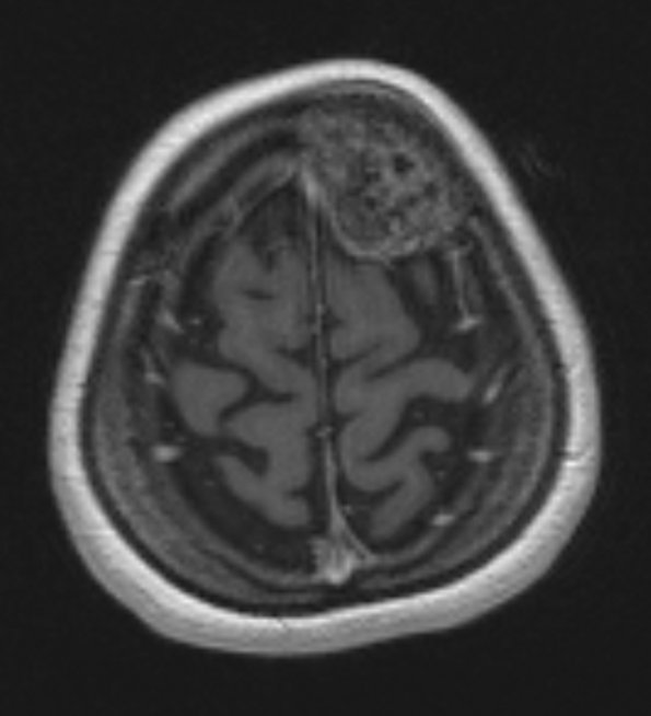
Washington University Experience | VASCULAR | Hemangioma | 12A1 Hemangioma (Case 12) T1 with contrast - Copy
Case 12 History ---- The patient is a 48 year old woman with a skull mass reportedly resected in 2002 by a general surgeon with pathology reportedly consistent with an osteoma. She has a well-circumscribed lesion in the left frontal skull region just eccentric to the midline that indents but does not involve the dura. Clinical and radiological differential was consistent with an aggressive hemangioma versus calvarial plasmacytoma. ---- The lesion is mildly hyperintense compared to adjacent cerebral cortex in this T1-weighted with contrast image.

