Table of Contents
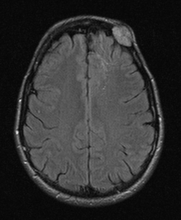
Washington University Experience | VASCULAR | Hemangioma | 5A1 Hemangioma (Case 5) FLAIR 1 - Copy
Case 5 History ---- The patient is a 45 year old woman with enlarging skull mass in the left frontal area. She underwent a biopsy of the lesion at age 43, and states that it has increased in size four fold since then. Operative procedure: Craniotomy. Diagnosis: Hemangioma, Cavernous ---- 5A1-3 MRI studies: Slightly hyperintense mass shown by FLAIR examination. The underlying cortex and white matter appear abnormal.

