Table of Contents
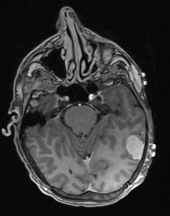
Washington University Experience | VASCULAR | Hemangioma | 7A1 Hemangioma (Case 7) T1 MPRAGE 1 - Copy
Case 7 History ---- The patient is a 32-year-old man with Klippel-Trenaunay-Weber (KTW) syndrome who presented with altered mental status. KTW syndrome is associated with large cutaneous hemangiomata or complex capillary-venous malformations, in some cases with hypertrophy of the related bones and soft tissues. A missense mutation in PIK3CA has been described as a somatic mosaic in some cases. The current patient has a past history of GI bleeding secondary to hemangiomas. Brain MRI shows a left sided extra-axial dural based contrast enhancing lesion. Clinical diagnosis: Meningioma versus hemangioma. Operative procedure: Craniotomy with resection of mass. ---- The hemangioma is hyperintense in MPRAGE.(7A1), T1 weighted image with contrast (7A2) and T2 weighted image with contrast (7A3)

