Table of Contents
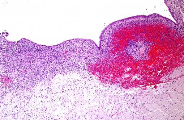
Washington University Experience | VASCULAR | Hemorrhage - Neonatal | 4B1 Hemorrhage, IVH & SEGM (Case 4) H&E 1
Low magnification image of a typical SEGM hemorrhage which is largely confined to the matrix with a few small erosions of the ependyma, as shown in image 4B2 (H&E).

