Table of Contents
Washington University Experience | VASCULAR | Herniation, uncal | 11A1 Herniation, uncal (PCA, Kernohan's (Case 11)
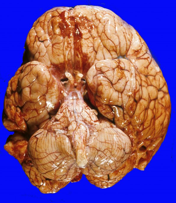
11A1,2 Grooved unci are seen on both sides although the discoloration of the left uncus is most impressive. Notice the unusual necrotic areas on the orbital surfaces of the frontal lobe, an appearance which was also seen in the temporal and occipital lobes; all of these lesions have a microscopic appearance of infarction with an inflammatory and microglial response. In this old case there was a suspicion for the presence of HSV-I encephalitis but inclusions were not identified by classical histology or electron microscopy and material for IHC and ISH is no longer available. There was no history of trauma. I have included this case because the uncal herniation findings are so striking.

