Table of Contents
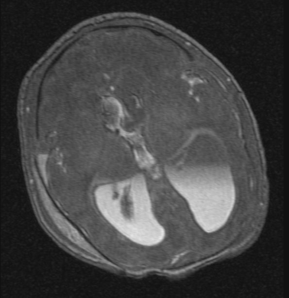
Washington University Experience | VASCULAR | Hydrocephalus, Post-Hemorrhagic | 3A1 Hydrocephalus IVH (Case 3) cow angio - Copy
A series of MRI examinations were performed on day of life 6 which showed marked ventriculomegaly, subependymal blood clot along the right choroid plexus and within both frontal horns, intraventricular hemorrhage extending into the third and fourth ventricles, and blood in the parenchyma along the right frontal horn. A small amount of subarachnoid blood was noted in the cortical sulci, bilaterally. Subdural hemorrhages were noted around the occipital poles, temporal poles, and along the interhemispheric fissure. Subgaleal hemorrhage was noted at the vertex.

