Table of Contents
Washington University Experience | VASCULAR | Hypoxia-Ischemia, fetal-neonatal | White Matter | 10C1 H-I, HLH, neonatal 34wk & 6wk (Case 10) A_4
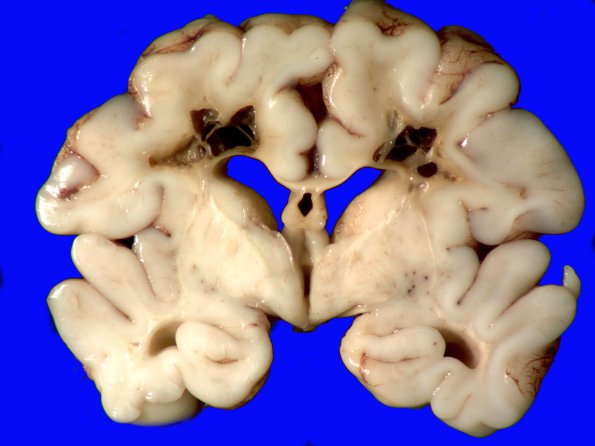
10C1-3 This coronal section was thought to represent a grade IV germinal matrix hemorrhage but, other than the orange stained walls, there was very little hemosiderin residua. The periventricular cysts are more consistent with periventricular necrosis, although scattered macrophages with hemosiderin are present within the cyst lining and may represent removal of much of the hemorrhagic residua. There were likely contributions by arterial and venous processes contributions with efficient resorption of blood elements. It is common for premature brains to have both hemorrhagic and ischemic components.

