Table of Contents
Washington University Experience | VASCULAR | Infarct, Tract Degeneration, illustrative case | 1A6 Infarct, remote (4 years), (Case 1) 13
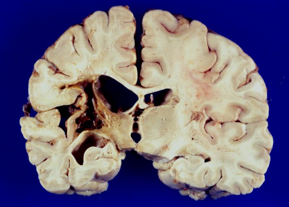
1A6-8 Multiple images of the cystic infarct at the level of the thalamus shows a loss of tissue with hydrocephalus ex vacuo involving the left lateral ventricle and temporal horn. The orange staining tissue reflects gliosis and scattered hemosiderin containing macrophages.

