Table of Contents
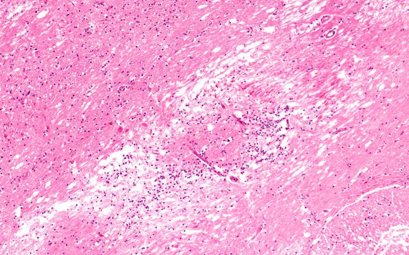
Washington University Experience | VASCULAR | Infarct - Acute | 4A2 Case 4 Pons N7 H&E 10X
4A2,3 These two sections of the basis pontis show red neurons (arrowhead, 4A3), axonal spheroids (arrow, 4A3) and PMNs infiltrating the perivascular spaces and parenchyma. Spheroids associated with longitudinal axons are prominent in this image. Axonal spheroids may develop very rapidly as axons begin to swell with transported materials as soon as hypoxia/ischemia begins whereas neurons take hours to actively develop H&E discernable eosinophilic neuronal necrosis. (H&E)

