Table of Contents
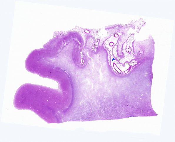
Washington University Experience | VASCULAR | Infarct - Remote | 2B Infarct Edge, Old (Case 2) b copy
A whole mount image of the margin of the infarct with normal brain. Note the cystic loss of gray matter with relative sparing of cortical layer 1 (arrowhead) and pallor of the underlying white matter. (H&E)

