Table of Contents
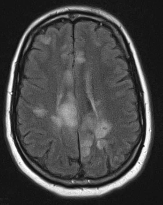
Washington University Experience | VASCULAR | Infarct - Subacute | 6A1 Infarcts, acute to chronic (Case 6) FLAIR 3 - Copy - Copy
Case 6 History ---- The patient was a 59-year-old man with a history of diffuse large B-cell lymphoma, hypertension, diabetes, hyperlipidemia, and a stroke in October of 2011. A head CT in August 2011 showed a large right parietal hematoma and he died two days later. Microscopy showed two resolving infarcts separate from the hematoma consisting of volume loss, sheets of macrophages (including a few hemosiderin laden macrophages) and exuberant reactive astrocytosis but no neoplastic cells.
6A1,2 MRI examination of the patient showed numerous lesions spread throughout the CNS as seen in FLAIR (16A1) and T2-weighted non-contrast enhanced (16A2) images.

