Table of Contents
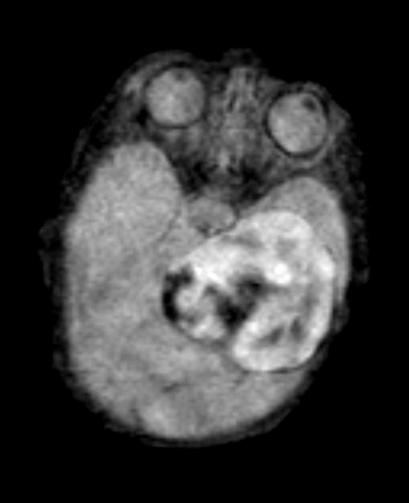
Washington University Experience | VASCULAR | Intravascular Papillary Endothelial Hyperplasia (Masson Tumor) | 2A1 PEH 7day old (Masson Tumor, Case 2) FLAIR - Copy
Case 2 History ---- The patient is a seven day old infant girl born at 35 weeks gestational age with an intracranial, extra axial mass located in the left middle fossa and extending into the posterior fossa as well. MRI studies show a partially cystic and solid, variably enhancing mass lesion. Clinical diagnosis: Teratoma vs neuroblastoma. Operative procedure: Biopsy of left middle cranial fossa mass. ---- MRI studies: The FLAIR (2A1), T1-weighted (2A2), TOF fi3D (2A3) and T2-weighted (2A4) images typically show a variegated pattern. ---- 2A1 FLAIR images of the lesion are hyperintense compared to brain parenchyma.

