Table of Contents
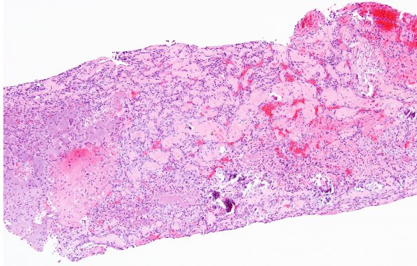
Washington University Experience | VASCULAR | Intravascular Papillary Endothelial Hyperplasia (Masson Tumor) | 2B1 Intravascular Papillary Endothelial Hyperplasia (Case 2) H&E 1
2B1-4 These images are those of a solid growth pattern with central hyalinized papillary cores of vascular elements lined by plump epithelioid cells, rare small hemosiderin deposits and patchy calcifications (H&E) Occasional mitoses are noted, although the cytologic features are bland.

