Table of Contents
Washington University Experience | VASCULAR | Intravascular Papillary Endothelial Hyperplasia (Masson Tumor) | 4A1 Meningioma, Radio Rx & Intravascular PEH (Case 4) H&E 33
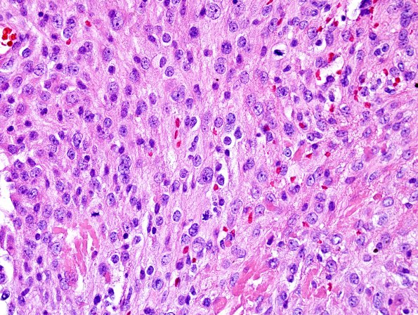
4A1-3 Sections of the resected left frontal parasagittal tumor show an atypical WHO Grade II meningioma with extensive post-treatment changes that include necrosis and vascular hyalinization. There are up to 7 mitotic figures in 10 HPF. Additional atypical features include sheeted architecture, hypercellularity, and many nuclei with prominent macronucleoli. (H&E)

