Table of Contents
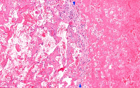
Washington University Experience | VASCULAR | Intravascular Papillary Endothelial Hyperplasia (Masson Tumor) | 5B3 Masson Tumor (Case 5) B1 H&E 10X 1 copy
In this area the right portion of the image consists of clot and fibrin which borders a transitional region (arrowheads) and a left portion of the field with well established PEH. (H&E)

