Table of Contents
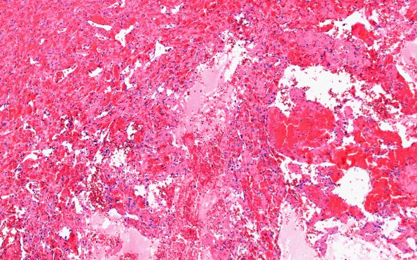
Washington University Experience | VASCULAR | Intravascular Papillary Endothelial Hyperplasia (Masson Tumor) | 5B4 Masson Tumor (Case 5) H&E 10X
5B4-8 Higher magnification images of the PEH portion of the specimen shows papillary structures lined by flattened spindled cells (shown later in immunohistochemical analysis to be endothelial cells). (H&E)

