Table of Contents
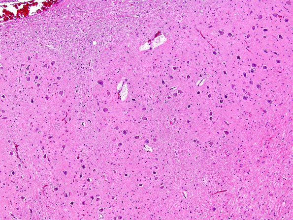
Washington University Experience | VASCULAR | Telangiectasis | 5H3 Telan&OliveHT (Case 5) H&E 10X NL area
5H3,4 Same magnification (10X) comparisons of dorsal (normal, 5H3) and ventral (hypertrophic vacuolated, 5H4) images of the IOH shows marked differences in size of neurons and vacuolated “mulberry” appearance (H&E)

