Table of Contents
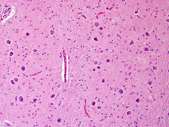
Washington University Experience | VASCULAR | Telangiectasis | 5H5 Telan&OliveHT (Case 5) H&E 20X NL ventral part.jpg
5H5,6 Same magnification (20X) comparisons of dorsal (normal, 5H5) and ventral (hypertrophic vacuolated, 5H6) images of the IOH shows marked differences in size of neurons and vacuolated appearance as well as striking atypical gemistocytic astrocytes (H&E)

