Table of Contents
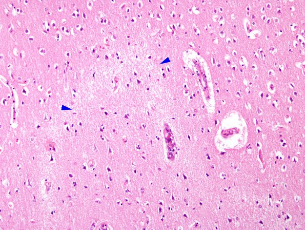
Washington University Experience | VASCULAR | Thrombotic Thrombocytopenic Purpura (TTP) | 3A1 TTP (Case 3) H&E 3 A
The cerebral hemispheres were symmetrical and appear normal to eye and palpation. Coronal sections through the cerebral hemispheres reveal no evidence of asymmetry or petechial hemorrhage. The centrum semiovale, convolutional white matter, basal ganglia, corpus callosum, thalamus, septum pellucidum, fornixes, and internal capsules are not remarkable without hemorrhagic foci noted. ---- 3A1-3 Microscopically, however, fibrin-platelet thrombi in arterioles and capillaries were widespread. Ischemic neuronal changes were, likewise, diffuse. An area of infarction is shown between two arrowheads (3A1) 3A1,2 Fibrin thrombi have resulted in microinfarcts (arrowheads, 3A1) characterized by pallor, eosinophilic neuronal necrosis, edema and microthrombi these changes were most obvious in the left temporal and parietal cortices, hippocampus, basal ganglia and pons. Fibrin-platelet thrombi were also found in the heart, lungs, kidneys, liver, adrenals and pancreas. (H&E)

