Table of Contents
Washington University Experience | VASCULAR | Vasculature, Ultrastructure | 1A1 Venule with Elastic, muscle (Case 1) 015 - Copy copy
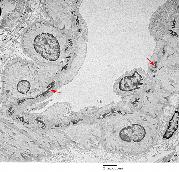
1A1-4 Ultrastructure of well-preserved venule from a muscle biopsy. ---- This low magnification image shows a small venule with a non-continuous single layer of elastin (arrows). Although thick internal elastic membranes are typically seen in arterial/arteriolar size vessels, smaller amounts are not uncommon in venules. (electron micrograph)

