Table of Contents
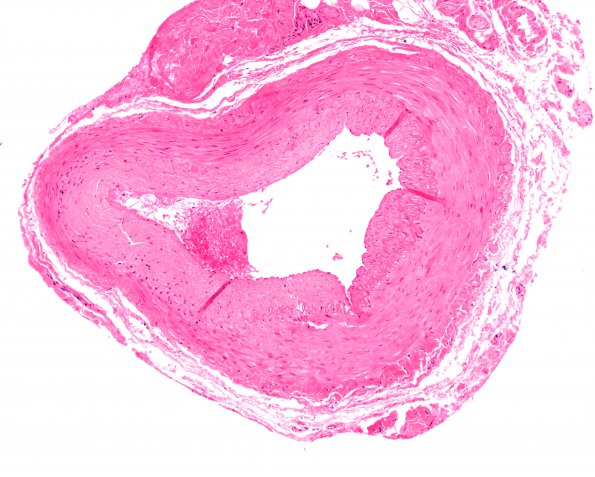
Washington University Experience | VASCULAR | Vasculitis - Giant Cell Arteritis | 5A1 Vasculitis, temporal artery, healed (Case 5) 10X
Case 5 History ---- The patient is a 72 year old man with a clinical diagnosis of temporal arteritis. Operative procedure: Biopsy of left temporal artery. There is patchy marked intimal thickening with focal areas of myxoid degeneration and loss of the internal elastic lamina. However, no definite lymphocytic infiltrate, epithelioid macrophages, or multinucleated giant cells were seen and the diagnosis of healed temporal arteritis was made. ---- 5A-D I have examined the same region of this cross section of the temporal artery stained for a variety of constituent elements at 10X, 20X and 40X. 5A1-3 There is little abnormal in this H&E stained series of magnifications although it appears somewhat hypocellular in smooth muscle areas which will be shown abnormal in latter stains (H&E)

