Table of Contents
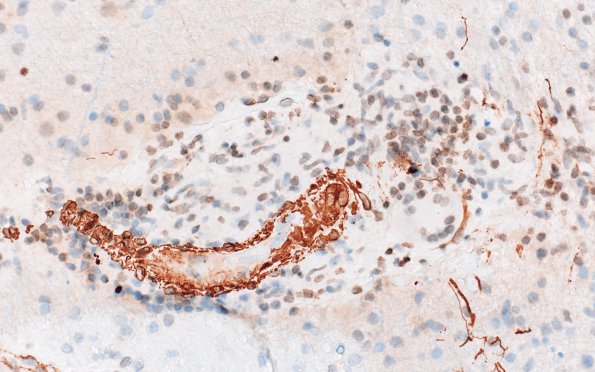
Washington University Experience | VASCULAR | Vasculitis - PACNS | 16C2 Granulomatous Angiitis (Case 16) A2 SMA 40X
Higher magnification of image #16C1. (SMA IHC)
Not shown:
GFAP highlights reactive astrocytes and background glial processes. CD68 shows perivascular accumulation of macrophages, as well as activated microglia and scattered parenchymal macrophages. Elastic stain does not identify vessels with elastic laminae. GMS and AFB stains are negative for fungal organisms and acid fast bacilli,

